Did You Know Your Oxygen Costs? Discover How Much You Could Save!
Discover how much you could save on oxygen costs with our insightful guide. Uncover potential…
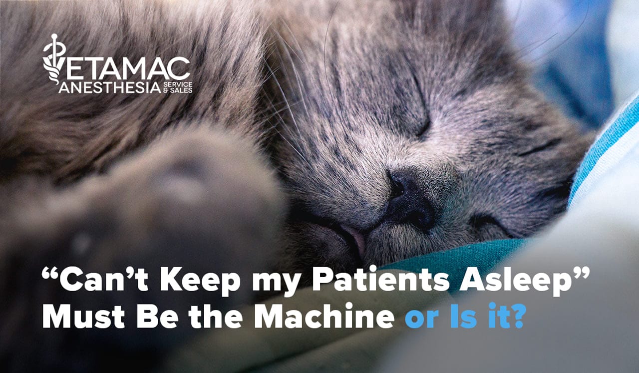
This is the most common complaint Vetamac service technicians hear from their veterinary clients. The most common component of the machine that receives the blame is the vaporizer. But a retrospective look at all of the 2010 Vetamac service records revealed that only 3.3% of the vaporizers verified were likely to be causing problems that would make it difficult to keep patients anesthetized1. The other components of the machine are a more likely cause for this problem.
The other components to check are the sodalime canister, flush valve, leaking domes, breathing circuits and rebreathing bags. The sodalime canister is one of the most common components for large leaks that make it difficult to keep patients anesthetized even on 5%. The canister gasket can become caked with soda lime dust and granules making it impossible to create a tight seal and therefore causing a large leak. Gaskets need to be cleaned every time the soda lime is changed. Also make sure the dust and granules are removed from the anesthesia machine’s breathing head where the canister is placed.
If the flush valve is leaking it will dilute the anesthetic gas in the breathing circuit and make it impossible to deliver a high enough concentration of anesthetic to keep patients anesthetized. The way to test the machine for a leaky flush valve is as follows. Place a breathing circuit and rebreathing bag on the machine and close the pop-off. Plug the end of the breathing circuit, pressurize the circuit to 20cm H20 by using the flush valve, turn off all oxygen flow and watch the manometer for a few seconds. If the flush valve is leaking the pressure will continue to rise. A service technician will be able to replace the flush valve. This pressure test will also test the breathing circuit and rebreathing bag for leaks.
So if the anesthesia machine passes the pressure test and/or the Vetamac service technician cannot find a leak, now what? Now the problem can be anesthesia techniques. One area to consider is the appropriate size and placement of the endotracheal tube (ET).
A common problem with the use of the endotracheal tube is the size. It is easy to use too small of an ET tube and then fill the cuff with a large (> 6ml) amount of air. Too small of a ET tube will increase the patient’s breathing and make it difficult to inspire a normal tidal volume. The Hagen-Poiseuille Law states that if the diameter of the airway is reduced then the resistance to gas flow will increase by a factor of four (McDonell and Kerr 2007). For example, if the tube selected is half of the normal diameter of the trachea, resistance to gas flow will increase by a factor of sixteenfold2.
Choosing the appropriate size ET tube is most challenging when it comes to veterinary patients. Due to the variety of breeds and sizes of the small animal patients it requires experience and knowledge to decide on the appropriate size. The ideal size ET tube should have the widest diameter that will pass comfortably into the trachea. The ET tube should never be forced into the trachea and the patient should be relaxed and not whining while passing the tube. Another way to estimate the appropriate size is to gently palpate the trachea and then have three sizes available prior to intubation. Always check the cuff prior to use to assure it is not leaking.
Once the ET tube is placed into the trachea, connect the breathing circuit and turn on the oxygen and vaporizer. The patient at this time may need a few breaths to further anesthetize them and then the following steps can be taken. Palpate the distal end of the ET tube before inflating the cuff and tying the tube to the patient. Once the proper placement is established, secure the tube and check it for leaks. This is not checking the machine for leaks it is checking the seal between the ET tube and the trachea. If the cuff requires more than 6 ml of air then the tube is too small. Once in the surgery room, check the cuff again for leaks. It is not uncommon to have to add another 1 ml or less of air to the cuff at this time since the patient is in a deeper plane of anesthesia and the walls of the trachea have relaxed.
Having more than one of each size and various lengths of ET tubes is helpful when preparing for the variety of veterinary patients. The two most common styles of ET tubes are the low volume high pressure (LVHP) and the high volume low pressure (HVLP) cuffs. The difference is as the name implies, the LVHP requires a small amount of air to create a tight seal on a small area but it exerts a very high pressure on the tracheal wall. In contrast, the HVLP cuff requires a large amount of air to create a seal and it is distributed over a wider surface area on the tracheal wall. This will cause low pressure on the trachea. All tubes when overinflated and used for long procedures can cause pressure necrosis. A hugely overinflated cuff can also cause the diameter of the ET tube to collapse and restrict the airway.
References
Tips from Deb


Veterinary Specialist Articles